


Uterine Fibroids
World-Class Experts
We are the oldest and largest private radiology group in the Omaha/Council Bluffs area, offering patients world-class expertise in the areas of:
- Abdominal imaging
- Breast Imaging
- Musculoskeletal Imaging
- Neuroimaging
- Nuclear medicine/molecular medicine
- Pediatrics
- Trauma
- Vascular and interventional radiology
Reliable Results
The accuracy of your diagnosis largely hinges on three things:
1) the technology of your diagnostic machines,
2) the experience of the person performing your tests, and
3) the specialization and experience of the radiologist who reads your reports
We have you covered with our top-of-the line equipment and the most experienced radiology team in the area.
You're in good hands!
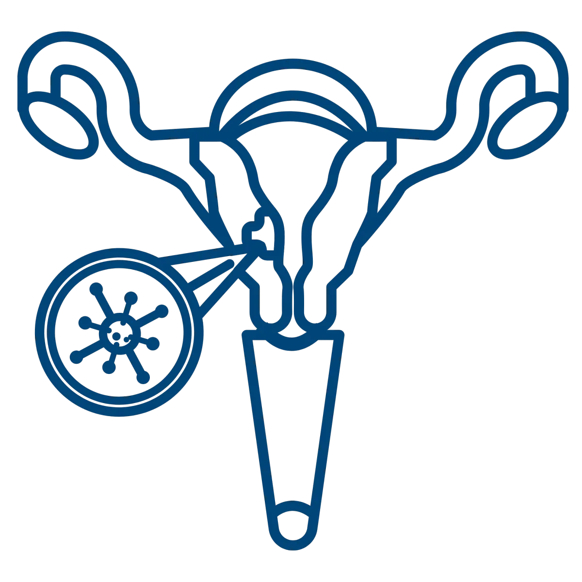
What are uterine fibroids?
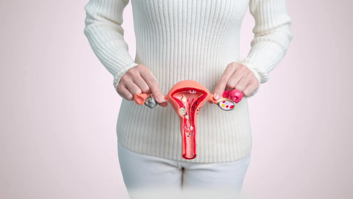
A uterine fibroid, also known as a leiomyoma or myoma, is a benign (non-cancerous) tumor that develops in the muscular wall of the uterus. These fibroids are composed of smooth muscle and connective tissue and can vary in size from small nodules to large masses. They can be located within the uterine wall, on its outer surface, or in the uterine cavity. Uterine fibroids can cause symptoms such as heavy menstrual bleeding, pelvic pain, frequent urination, or pressure in the lower abdomen, although some women may have no symptoms at all.
What are the symptoms of uterine fibroids?
Uterine fibroids can present with a range of symptoms, including:
Heavy menstrual bleeding
Pelvic pain or cramping
Frequent urination due to pressure on the bladder
Lower back pain
Pain during intercourse
Sensation of fullness in the abdomen or noticeable abdominal enlargement if the fibroids are large

However, some individuals may be asymptomatic, and fibroids may be detected incidentally during routine examinations or imaging.
How are uterine fibroids diagnosed?
Uterine fibroids are diagnosed using several methods:
Medical History & Physical Examination
The clinician gathers information about symptoms and performs a pelvic examination to assess abnormalities
Ultrasound
This imaging technique utilizes sound waves to visualize the uterus and identify the size and location of fibroids
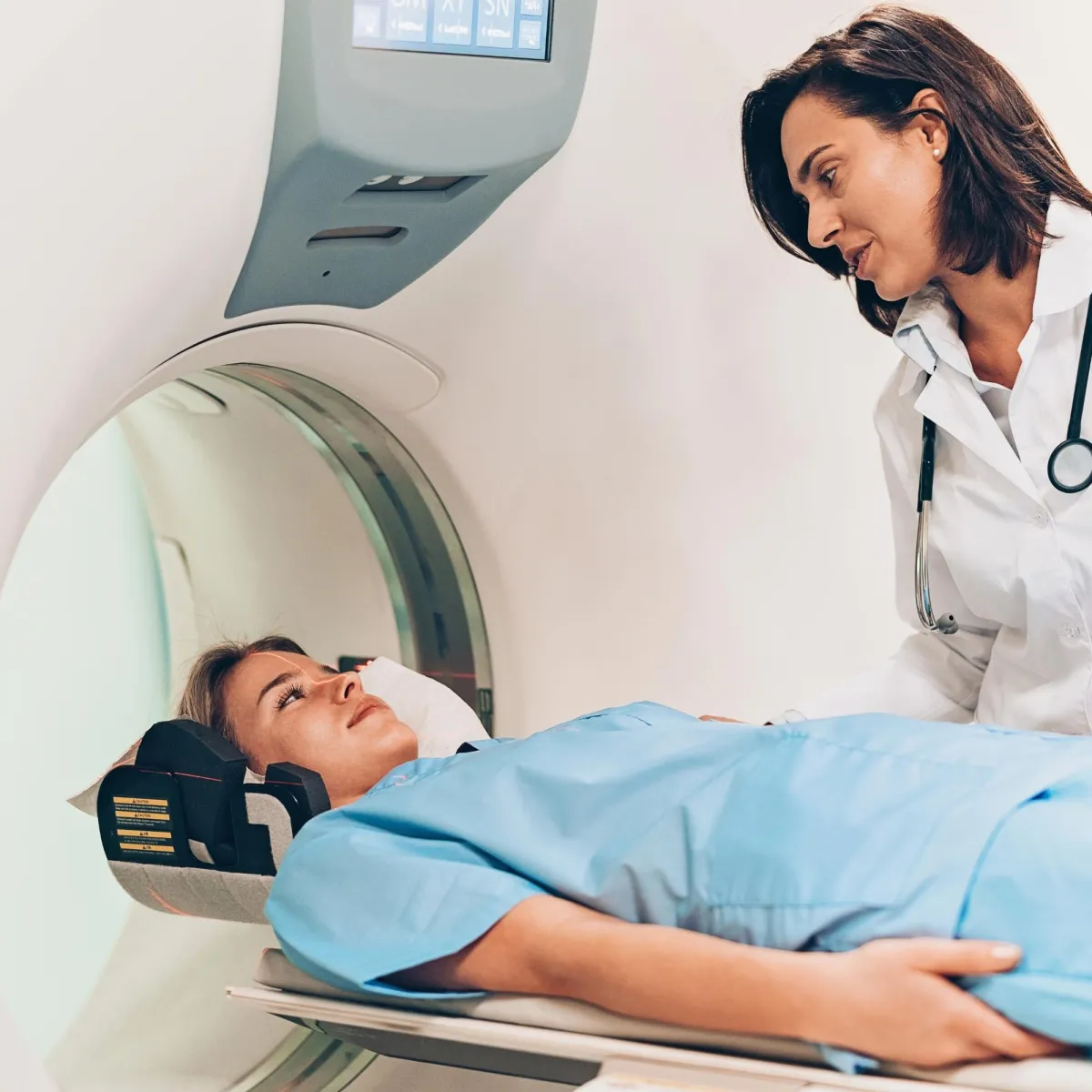
Magnetic Resonance Imaging (MRI)
Provides detailed and precise images of the uterus, especially useful for complex cases
Hysteroscopy
Involves inserting a thin, flexible scope through the vagina and cervix into the uterus to directly visualize and potentially biopsy fibroids within the uterine cavity
Sonohysterography
Also known as saline infusion sonography, this involves injecting a saline solution into the uterus during an ultrasound to enhance imaging of the uterine lining and fibroids
An MRI exam helps in selecting patients who should receive nonsurgical uterine fibroid embolization. Interventional radiologists interpret the MRI images to determine if a fibroid tumor can be embolized, detect alternate causes for the symptoms, identify conditions that could prevent the procedure, and avoid ineffective treatments. Patients should discuss their options with their gynecologist and interventional radiologist.
How MIC Treats Uterine Fibroids
Non-Surgical Procedures
Uterine Artery Embolization (UAE)
A minimally invasive procedure that blocks blood flow to the fibroids, causing them to shrink
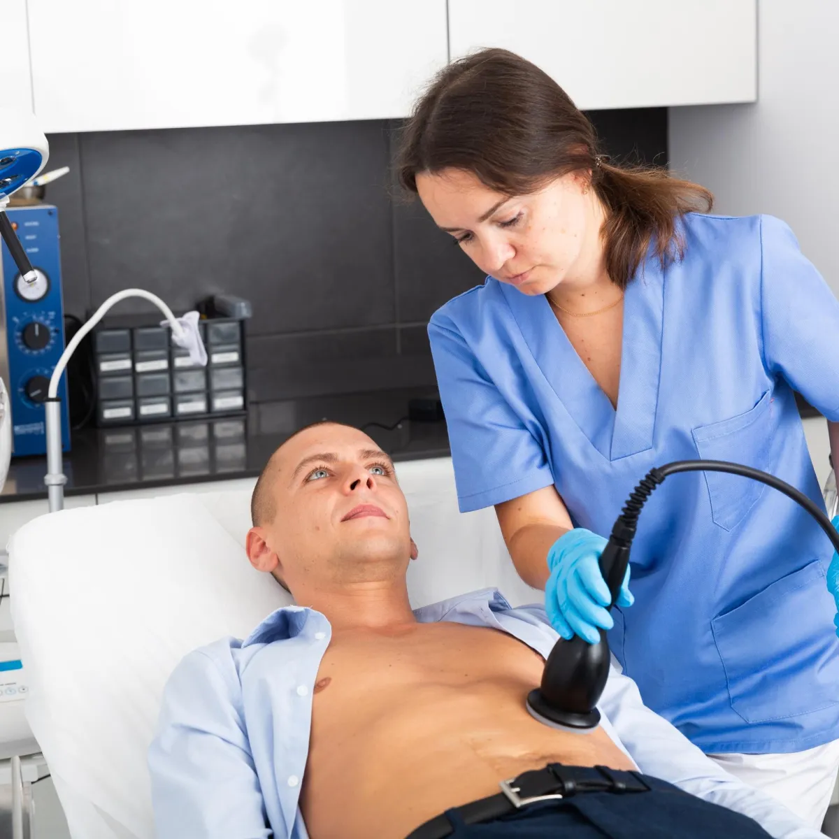
Focused Ultrasound Surgery (FUS)
Uses high-intensity ultrasound waves to destroy fibroid tissue
Surgical Procedures
Myomectomy
Surgical removal of fibroids while preserving the uterus, which can be performed via hysteroscopy, laparoscopy, or abdominal surgery
Hysterectomy
Complete removal of the uterus is considered when fibroids are severe or when other treatments are not suitable
Uterine fibroid embolization is a minimally-invasive surgical procedure performed by an interventional radiologist to treat conditions involving the presence of uterine fibroid tumors by blocking their blood supply and shrinking them.
Minimally Invasive Procedure Specialists utilize uterine fibroid embolization, or UFE, as a non-surgical treatment for fibroids. Non-surgical procedures like UFE are much less invasive, in most instances do not require a hospital stay and allow for quicker recovery. Learn more about UFE and call our office in Denver, CO to schedule your consultation. You can also request an appointment online. A member of our staff will reach back out to you to confirm the details of your appointment shortly after the request has been received.
About Your Procedure
Uterine Fibroid Embolization (UFE) is a minimally invasive procedure used to treat uterine fibroids. It involves inserting a catheter through a small incision and guiding it to the uterine arteries. Tiny particles are then injected to block blood flow to the fibroids, causing them to shrink and alleviate symptoms like heavy bleeding and pelvic pain. The procedure typically requires a short recovery time, with most patients resuming normal activities within a week, and it preserves the uterus.
Focused Ultrasound Surgery (FUS) is a non-invasive treatment for uterine fibroids. During the procedure, the patient lies on a treatment table while a machine uses high-intensity ultrasound waves to target and heat the fibroids. This heat destroys the fibroid tissue while preserving the surrounding healthy tissue. The treatment is guided by real-time imaging to ensure accuracy. Recovery is typically quick, with many patients returning to normal activities within a few days.
Uterine fibroid embolization takes about an hour. After the procedure, the patient is required to remain in a hospital setting overnight. Most women experience moderate to severe pain and cramping for several hours after having the procedure. On occasion, some may also experience fever and nausea. The majority of women are able to return to normal activity 7 to 10 days after the procedure and many resume light activities in a few days. Between 85 to 90 percent of women experience significant or complete relief of heavy bleeding and relief from pain.
Varicose
Veins
Pelvic
Pain
Non Healing Wounds
Plantar
Fasciitis
Uterine
Fibroids
Benign Prostatic Hyperplasia
Services


CT Scan
Minimally Invasive Therapies
Molecular Medicine


MRI
Ultrasound
Women's Imaging

M-F: 8:00am-5:00pm
Extended Hours Available
Upon Request

