


Mammography
Mammography, using the advanced Hologic 3Dimensions™ system, is a specialized X-ray imaging technique designed to detect and evaluate abnormalities in breast tissue. Hologic 3D mammography exams are more accurate than conventional 2D mammograms, detecting 20%-65% more invasive cancers. During the procedure, the breast is positioned between two plates in the mammography machine, which compresses the tissue to capture clear, detailed X-ray images from different angles. This compression is brief but necessary to ensure high-quality images and minimize movement. To prepare, avoid using deodorants, lotions, or powders on the breasts or underarms, as these can interfere with the images. It is also helpful to schedule the mammogram for a time when your breasts are less likely to be tender, typically not during menstruation.
World-Class Experts
We are the oldest and largest private radiology group in the Omaha/Council Bluffs area, offering patients world-class expertise in the areas of:
- Abdominal imaging
- Breast Imaging
- Musculoskeletal Imaging
- Neuroimaging
- Nuclear medicine/molecular medicine
- Pediatrics
- Trauma
- Vascular and interventional radiology
Reliable Results
The accuracy of your diagnosis largely hinges on three things:
1) the technology of your diagnostic machines,
2) the experience of the person performing your tests, and
3) the specialization and experience of the radiologist who reads your reports
We have you covered with our top-of-the line equipment and the most experienced radiology team in the area.
You're in good hands!
What is mammography?
Mammography is a specialized X-ray imaging technique designed for the early detection and diagnosis of breast abnormalities.
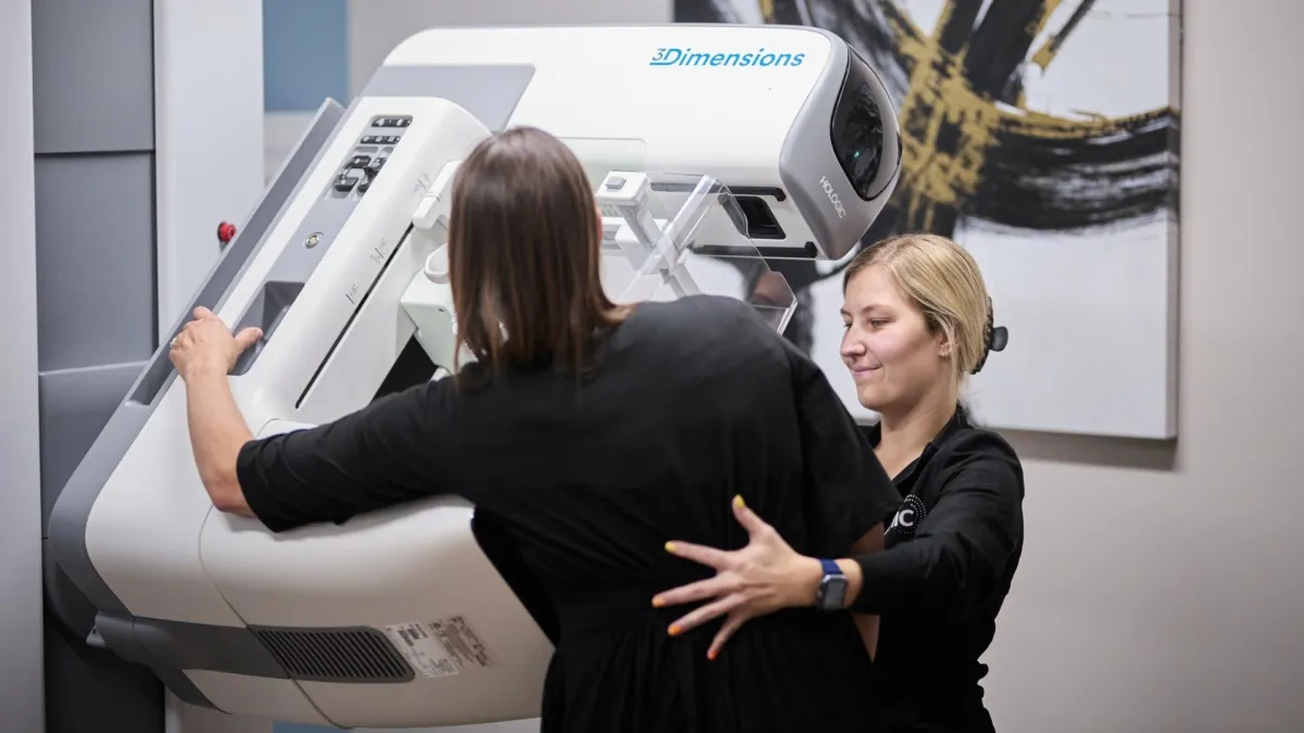
The procedure involves compressing the breast between two plates within a mammography machine to obtain high-resolution X-ray images from different angles. This compression is necessary to provide clear and detailed images while minimizing movement and radiation exposure. Mammography is a critical tool in screening for breast cancer, detecting tumors or other changes in breast tissue at an early stage when treatment options are often more effective. The results are analyzed by a radiologist, who provides a report to the referring physician for further evaluation and management.
In certain cases, an ultrasound of the breast is performed in conjunction with diagnostic mammography. This exam is non-invasive and uses sound waves, rather than radiation, to produce images. The ultrasound technologist will move a wand coated in gel over the breast tissue to further examine areas of concern. The images produced can provide greater clarity in the evaluation of the breast tissue.
Important things to know about mammography at MIC
Mammography is used to detect and diagnose breast abnormalities, including breast cancer, at an early stage. It helps in screening asymptomatic individuals and monitoring known conditions.
During a mammogram, the breast is positioned between two plates that compress it to obtain clear images. This compression is necessary to reduce motion and ensure the highest quality images, although it may cause temporary
discomfort.
The amount of radiation used in mammography is very low and within safety limits. If you have concerns about radiation exposure, discuss them with your healthcare provider. The procedure typically lasts only a few minutes.
The procedure may cause some discomfort due to the compression of the breast, but it is typically brief, lasting only a few minutes. The overall experience is generally well-tolerated, and the benefits of early detection often outweigh the temporary discomfort.
While mammography is a crucial tool for early cancer detection, it is important to understand that it is not perfect. False positives (indicating cancer when there is none) and false negatives (missing existing cancer) can occur. The benefits include early detection, which can lead to more effective treatment and better
outcomes.
After the mammogram, a radiologist will review the images and send a report to your healthcare provider, who will discuss the findings with you. If any abnormalities are detected, further tests or follow-up appointments may be
recommended.
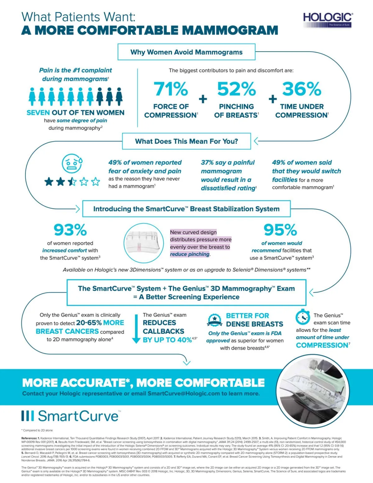
Experience the Softer Mammogram at MIC
MIC's state-of-the art equipment combines SmartCurve(TM) and Genius 3D Mammography(TM) technology, resulting not only in a more accurate exam, but a more comfortable one as well.
Learn more:
Frequently Asked Questions
Do I need a mammogram?
The decision to have a mammogram is a personal one and is dependent upon age, family history, the density of breast tissue, and discussion with your physician. Ultimately – it’s your decision – you do not need a recommendation from a physician for a screening mammogram. In terms of age, research clearly shows that annual mammography and breast cancer screen should start at age 40 and it is strongly correlated with significant improvement in survival rates of breast cancer.
How to Prepare for Your Mammogram
Please alert our staff when scheduling if you have had prior mammograms at other facilities, so that we can obtain the images prior to your appointment for comparison. This will ensure there is no delay in your exam being interpreted. Do not apply deodorants, antiperspirants, powders, or lotions to your underarm or breast area on the day of the exam. These products can interfere with the clarity of the images. Wipes are provided to remove these products if you forget, or unable to comply with this request. You will be asked to undress from the waist up, a robe or gown will be provided that opens in the front. Wearing a top and bottom is more convenient than a one-piece outfit. It is advisable to avoid scheduling the mammogram during times when breast tenderness can be heightened. One week following your period is usually best. This can help minimize discomfort during the compression. Also, inform the technologist if you have a history or breast cancer, breast implants, a history of breast surgery, or if you are pregnant or breastfeeding. Adhere to any additional instructions provided by your healthcare provider or the imaging facility. This might include details specific to your medical history or the type of mammogram being performed.
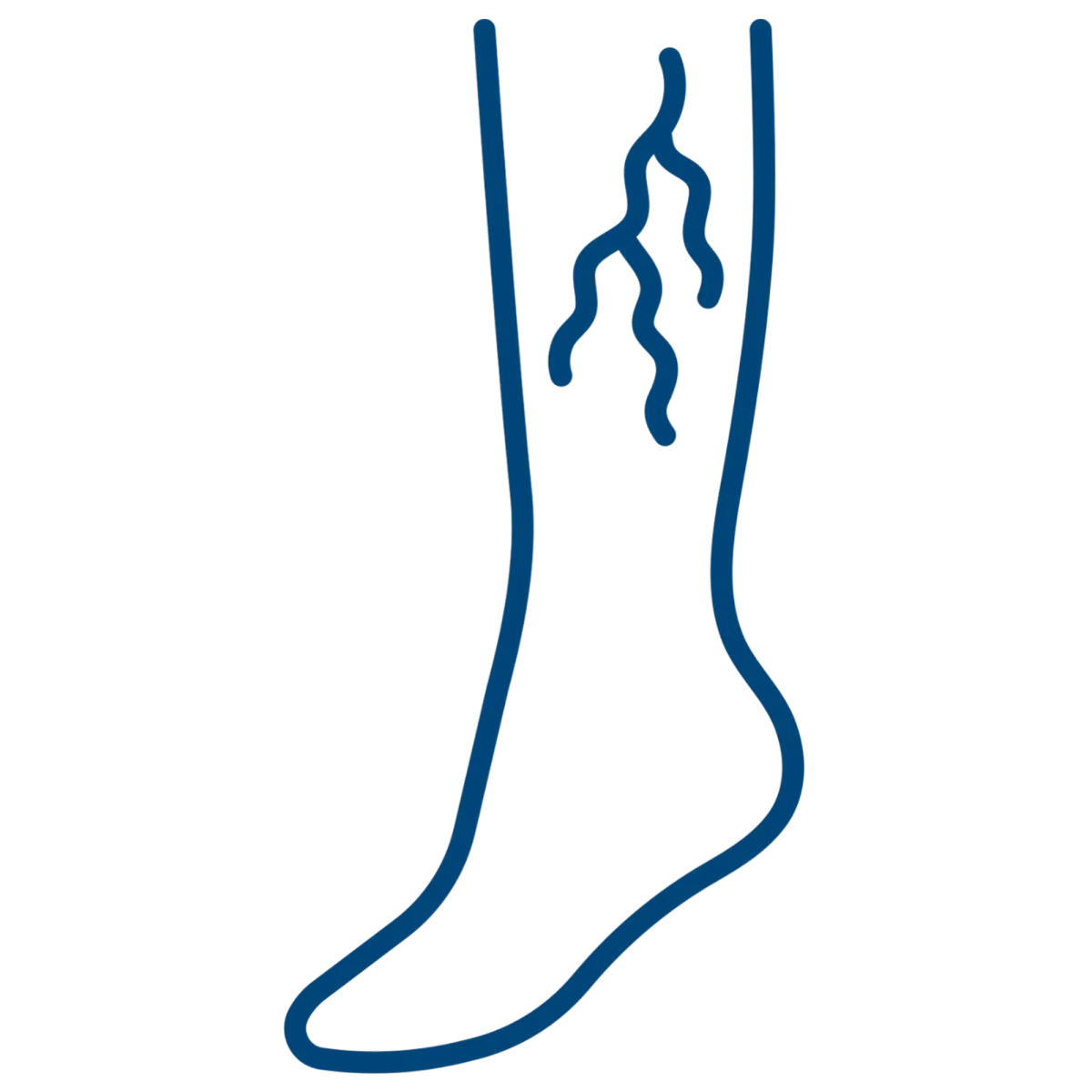
Varicose
Veins
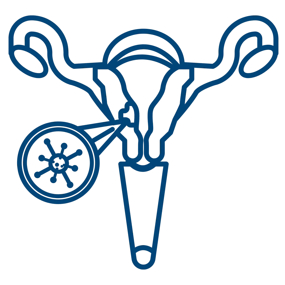
Pelvic
Pain
Non Healing Wounds
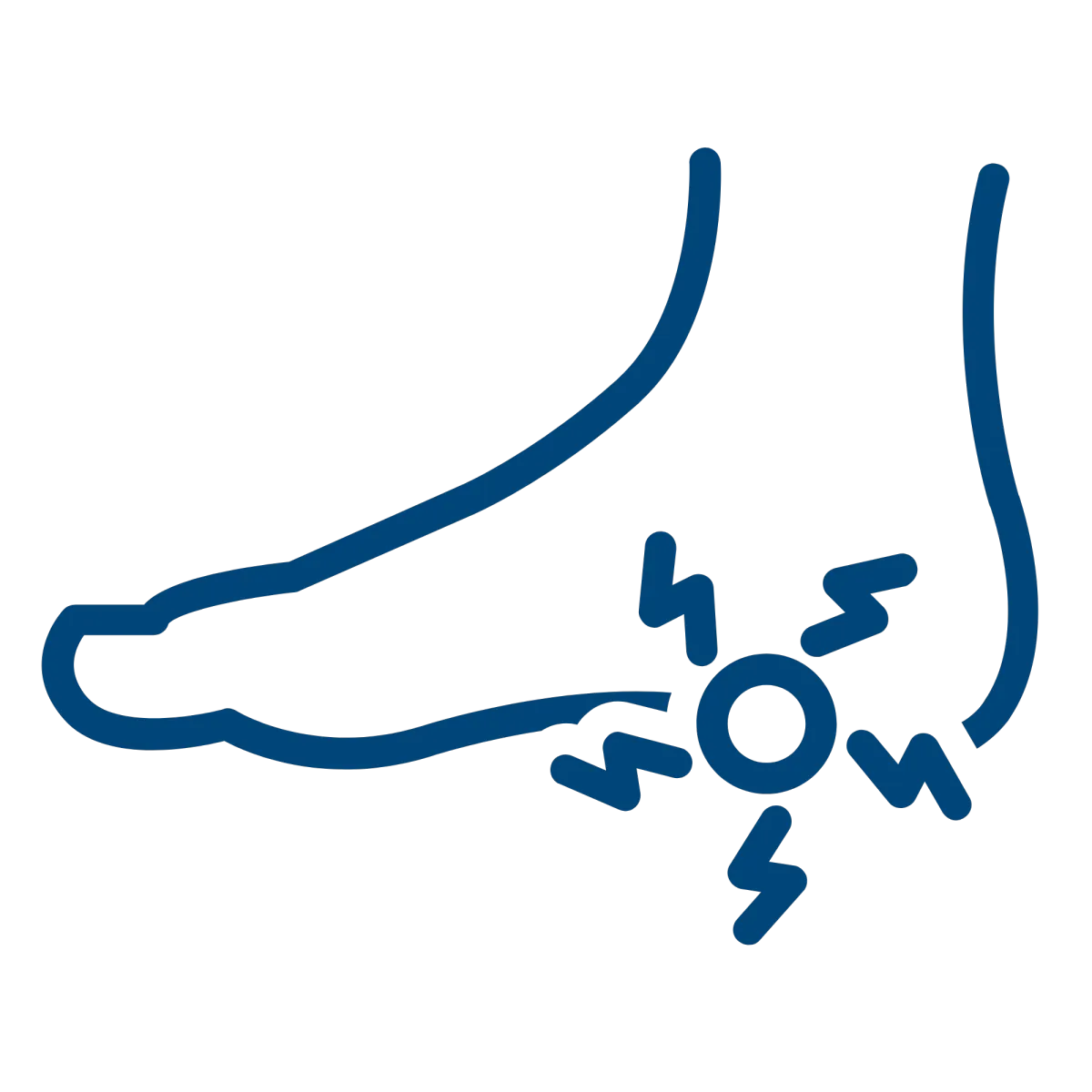
Plantar
Fasciitis
Uterine
Fibroids
Benign Prostatic Hyperplasia
Services


CT Scan
Minimally Invasive Therapies
Molecular Medicine


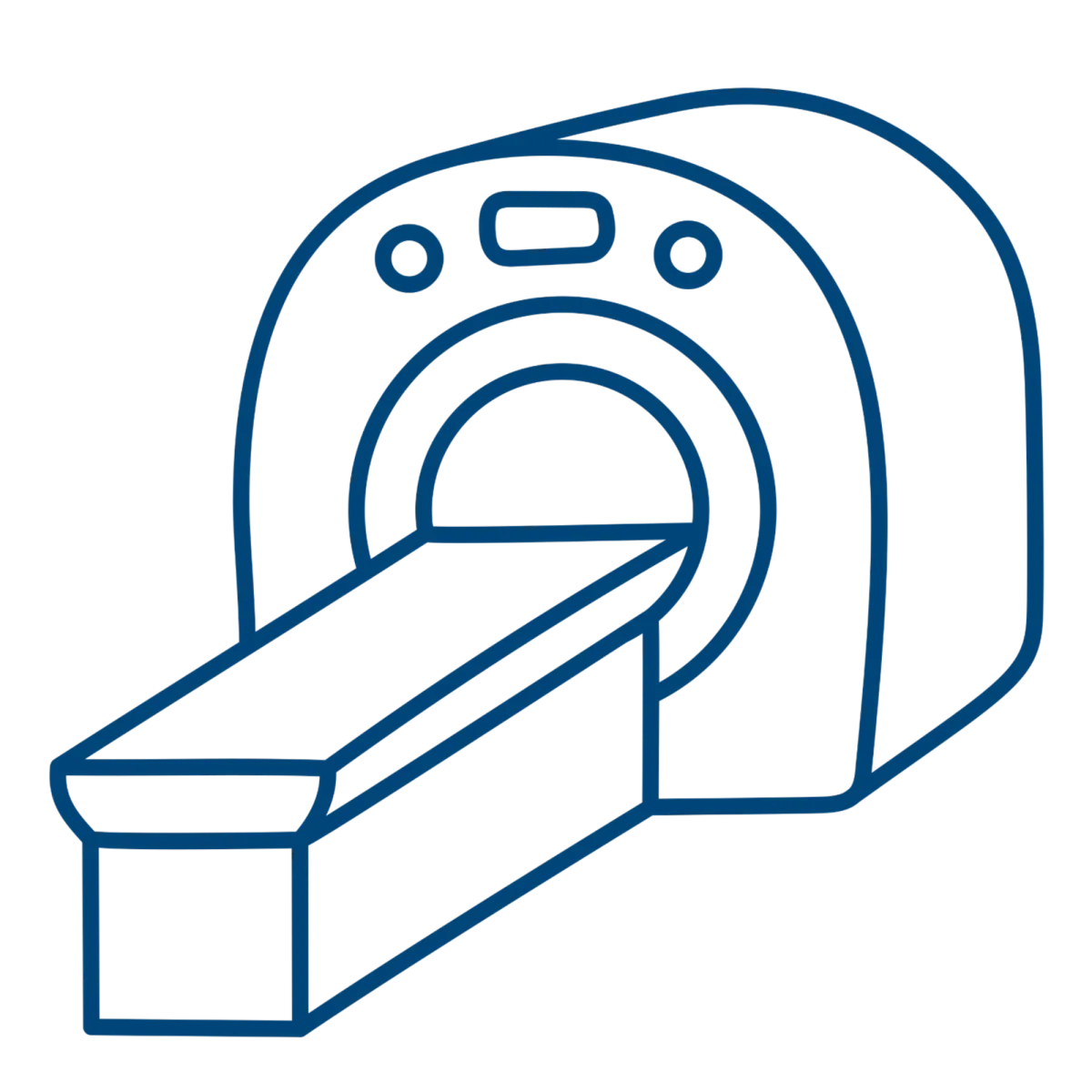
MRI
Ultrasound
Women's Imaging

M-F: 8:00am-5:00pm
Extended Hours Available
Upon Request

