


DEXA Bone Scan
A DEXA (Dual-Energy X-ray Absorptiometry) scan is a diagnostic imaging test used to measure bone density and assess the risk of osteoporosis and fractures. During the procedure, you lie on a table while a machine scans your body using low-dose X-rays to produce detailed images of bone. Typically focusing on the spine and hip. The scan is quick, typically lasting about 10 to 20 minutes, and involves minimal radiation exposure. A quantitative formula is applied to the images to determine your bone mass density and assess your risk for fracture. To prepare, wear loose, comfortable clothing and remove any metal objects that could interfere with the scan. Inform the technician if you have had any recent X-rays or other imaging studies, as this may affect the results.
World-Class Experts
We are the oldest and largest private radiology group in the Omaha/Council Bluffs area, offering patients world-class expertise in the areas of:
- Abdominal imaging
- Breast Imaging
- Musculoskeletal Imaging
- Neuroimaging
- Nuclear medicine/molecular medicine
- Pediatrics
- Trauma
- Vascular and interventional radiology
Reliable Results
The accuracy of your diagnosis largely hinges on three things:
1) the technology of your diagnostic machines,
2) the experience of the person performing your tests, and
3) the specialization and experience of the radiologist who reads your reports
We have you covered with our top-of-the line equipment and the most experienced radiology team in the area.
You're in good hands!
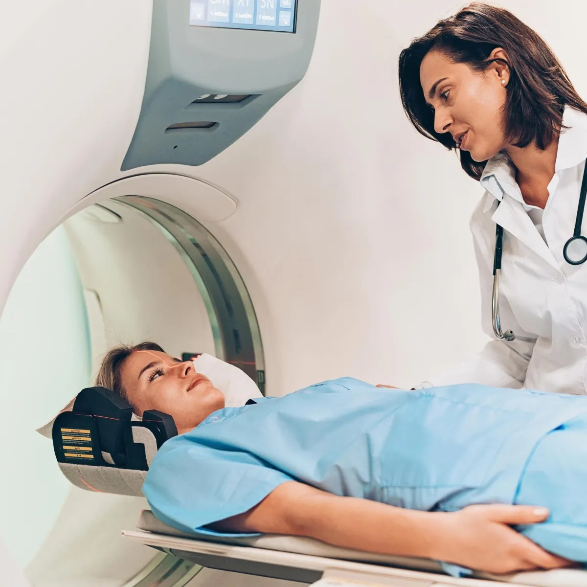
What is a DEXA bone density exam?
A DEXA scan is a specialized imaging technique used to measure bone mineral density (BMD) and assess the risk of osteoporosis and fractures.
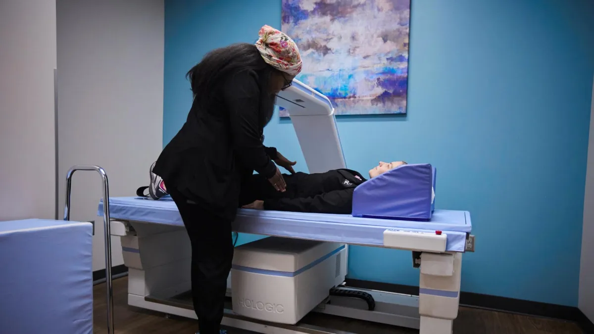
The procedure involves using two different X-ray beams with varying energy levels to evaluate bone density in key areas such as the spine, hip, and sometimes the forearm. By comparing the absorption of these X-ray beams, the DEXA scan provides detailed information about bone mass and strength. This non-invasive test typically requires the patient to lie on a table while a scanning arm passes over the body, emitting low-dose radiation. The results help healthcare providers diagnose bone-related conditions, monitor changes in bone density over time, and guide treatment decisions.
Bone Densitometry, also called dual-energy x-ray absorptiometry, DEXA or DXA, uses a very small dose of ionizing radiation to produce pictures of the inside of the body (usually the lumbar spine and hips) to measure bone loss. It is commonly used to diagnose osteoporosis, to assess an individual’s risk for developing osteoporotic fractures. DEXA is simple, quick and noninvasive. It’s also the most commonly used and the most standard method for diagnosing osteoporosis.
This simple 10-minute procedure compares your bone density to that of the average bone density of a 30-year old healthy woman.
Important Things to Know About a Bone Density Exam
The exam involves lying on a table while a scanning arm passes over specific areas of the body, such as the spine, hip, or forearm. The scan uses low-dose X-rays to evaluate bone density and typically takes about 10 to 20 minutes.
The procedure is non-invasive, painless, and involves minimal radiation exposure. The scan is quick, and you may be asked to lie still during the examination to ensure clear images.
DEXA is considered the gold standard for bone density measurement due to its accuracy, precision, and low radiation dose. It is valuable for diagnosing osteoporosis early and monitoring the effectiveness of treatment.
The results are analyzed by a radiologist who will provide a report to your healthcare provider. Your provider will discuss the findings with you and recommend any necessary follow-up actions or treatments based on your bone density measurements.
Frequently Asked Questions
How much does a bone density exam cost?
Medicare will pay for a bone density test as part of preventive screening every two years for women 65 or older and men 70 or older. Many insurance providers will cover the test under certain circumstances.
Can I get a bone density exam if I'm under the age of 65?
If you are over the age of 50 with a family history of osteoporosis or symptoms such as unusual fractures, you should consult with a physician to determine if you need a DEXA scan.
When will my results be available?
The radiologist will review the DEXA images and provide a diagnostic report that will be sent directly to your provider. The report is typically available to your provider within 24 hours. Many providers plan scheduled time to discuss results with their patients so you could check with their office to see when they will be available to review the information with you.
How to Prepare for a DEXA
Refrain from taking calcium supplements for at least 24 hours before the exam, as calcium can affect the accuracy of the results. It is best to wear loose, comfortable clothing without metal fasteners, such as zippers or buttons, which can interfere with the imaging process. You may be asked to change into a robe or gown provided by our facility. Notify the technologist about any recent X-rays or CT scans, as well as any medical conditions, particularly if you have had a recent fracture or are taking medications that affect bone density.
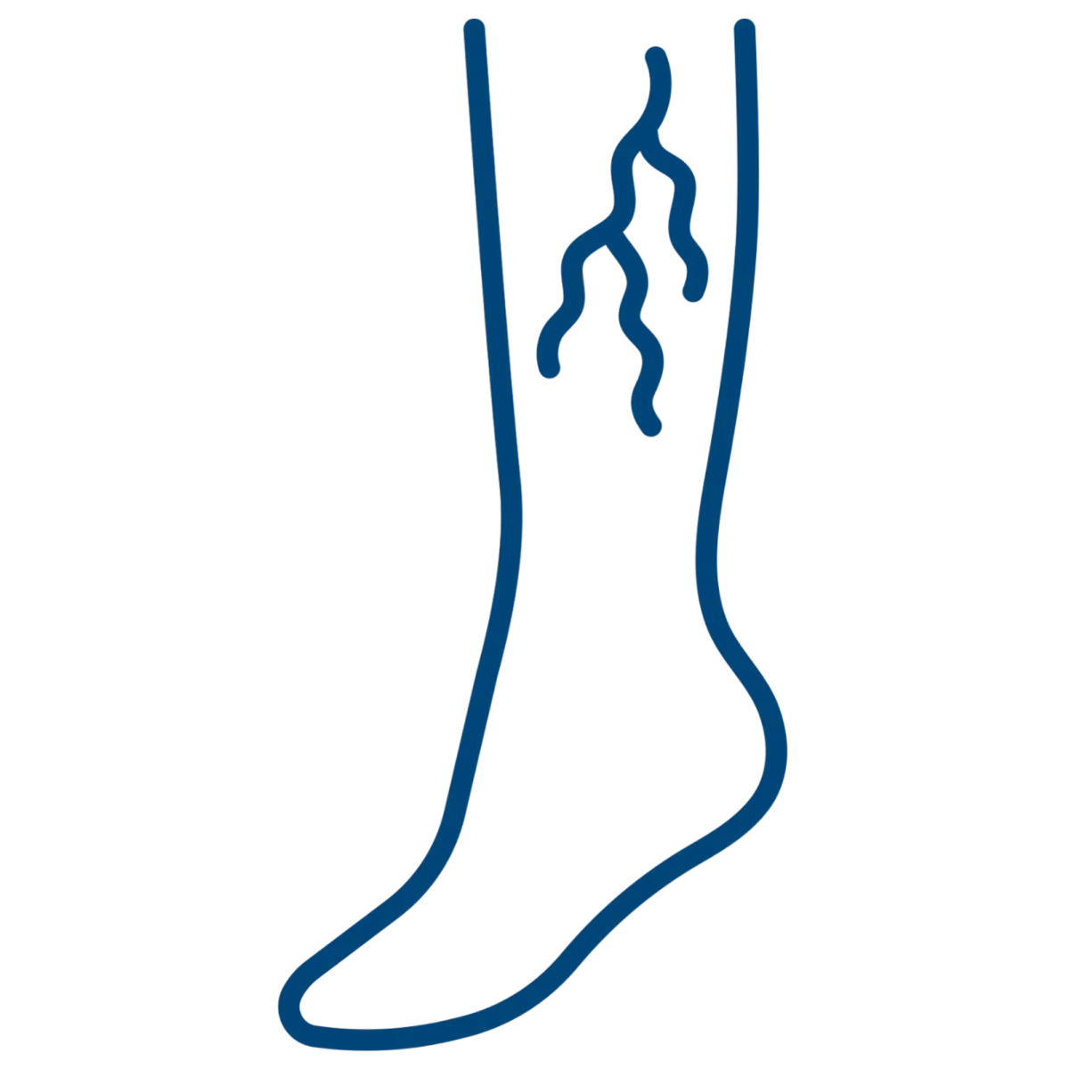
Varicose
Veins
Pelvic
Pain
Non Healing Wounds
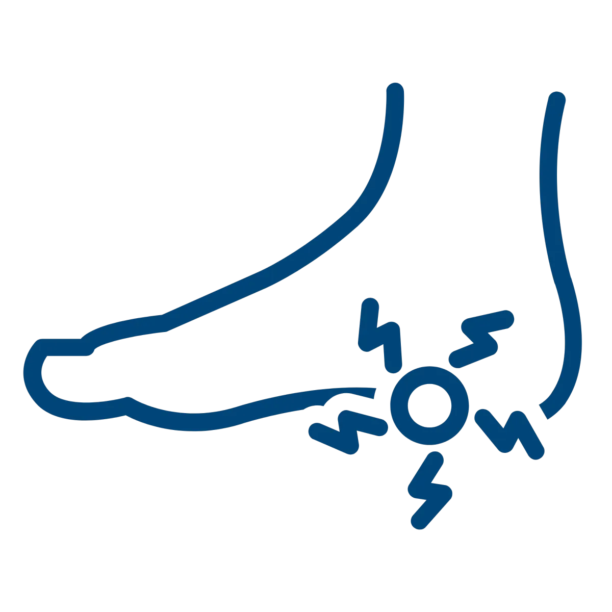
Plantar
Fasciitis
Uterine
Fibroids
Benign Prostatic Hyperplasia
Services


CT Scan
Minimally Invasive Therapies
Molecular Medicine


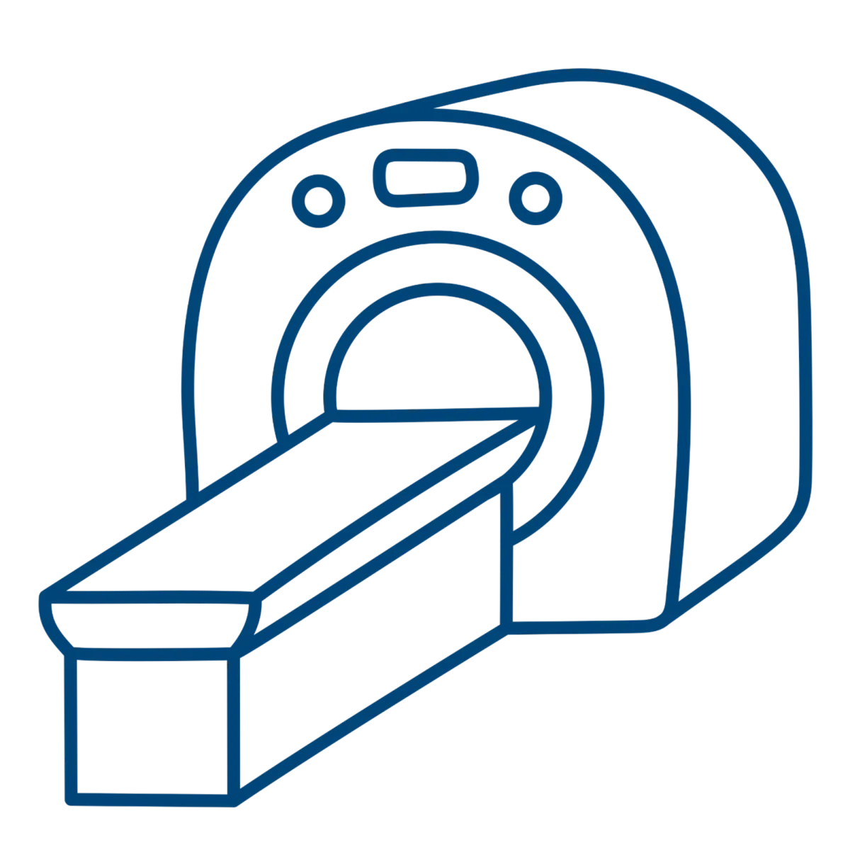
MRI
Ultrasound
Women's Imaging

M-F: 8:00am-5:00pm
Extended Hours Available
Upon Request

