


Breast Ultrasound
An ultrasound, or sonogram, is a diagnostic imaging technique that uses high-frequency sound waves to create real-time images of the inside of the body. During the procedure, a gel is applied to the skin, and a handheld device called a transducer is moved over the area of interest and slight pressure is applied to capture the sound waves reflected off internal structures. The images are then displayed on a monitor for evaluation. No special preparation is required for a breast ultrasound. The procedure is non-invasive, does not use radiation, and is generally painless.
World-Class Experts
We are the oldest and largest private radiology group in the Omaha/Council Bluffs area, offering patients world-class expertise in the areas of:
- Abdominal imaging
- Breast Imaging
- Musculoskeletal Imaging
- Neuroimaging
- Nuclear medicine/molecular medicine
- Pediatrics
- Trauma
- Vascular and interventional radiology
Reliable Results
The accuracy of your diagnosis largely hinges on three things:
1) the technology of your diagnostic machines,
2) the experience of the person performing your tests, and
3) the specialization and experience of the radiologist who reads your reports
We have you covered with our top-of-the line equipment and the most experienced radiology team in the area.
You're in good hands!
What is a breast ultrasound?
An ultrasound, also known as sonography, is a medical imaging technique that employs high-frequency sound waves to produce real-time images of internal structures within the breast tissue.

The process involves transmitting sound waves through a transducer, which is a handheld device that is moved over the skin after applying a layer of gel to the breast. These sound waves echo off tissues, creating visual images on a monitor that can be used to assess the tissue within the breast. It is very useful in accessing abnormalities in women with dense breast tissue. Ultrasound is widely utilized for evaluating organs, monitoring pregnancies, and guiding certain medical procedures due to its non-invasive nature and lack of ionizing radiation. The resulting images assist healthcare professionals in diagnosing conditions, planning treatments, and conducting follow-up assessments. Ultrasound is particularly useful in that it is often able to tell the difference between a fluid-filled mass like cysts (which are most often benign), and solid masses (these may need further evaluation to determine they are not cancer).
Frequently Asked Questions
How long will the Ultrasound take?
Most breast ultrasounds are completed in 20-30 minutes.
How to prepare for a breast Ultrasound
No special preparation is required for a breast ultrasound.
Varicose
Veins
Pelvic
Pain
Non Healing Wounds
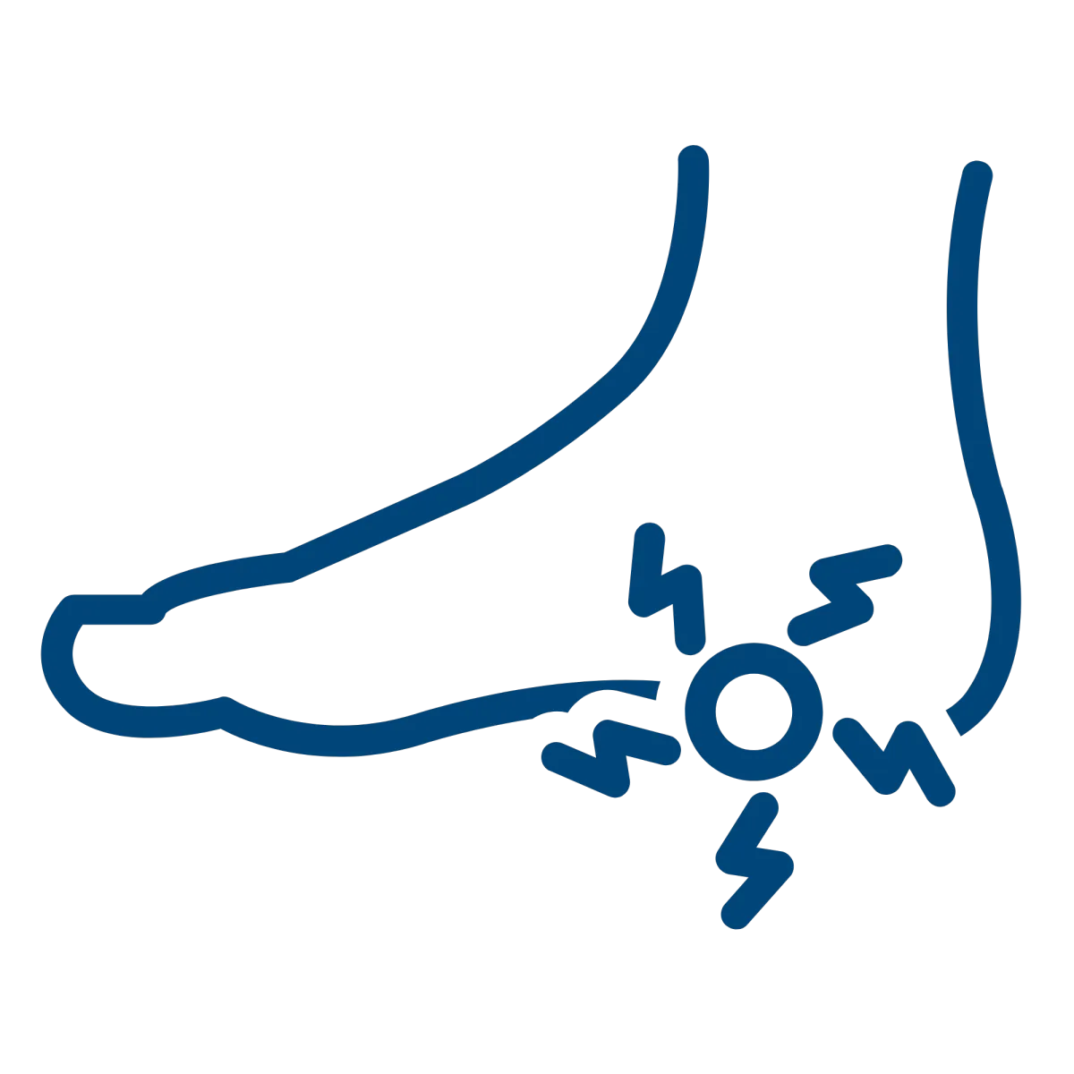
Plantar
Fasciitis
Uterine
Fibroids
Benign Prostatic Hyperplasia
Services
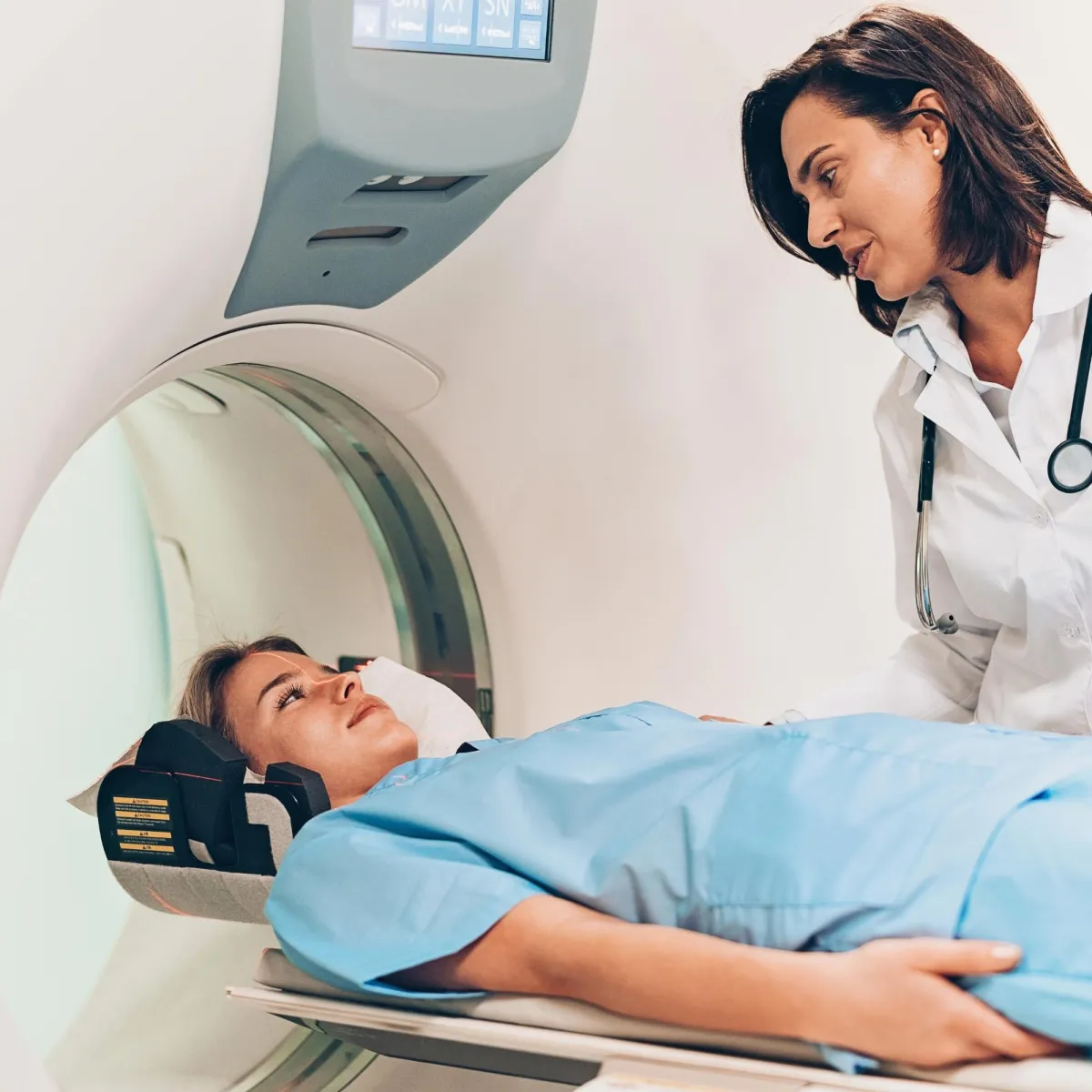


CT Scan
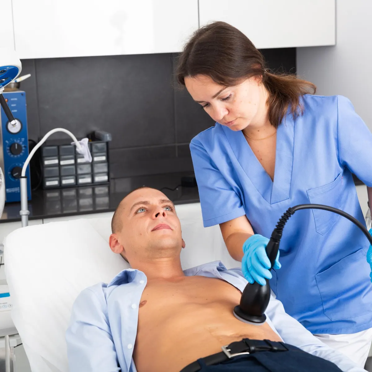
Minimally Invasive Therapies
Molecular Medicine


MRI
Ultrasound
Women's Imaging

M-F: 8:00am-5:00pm
Extended Hours Available
Upon Request

